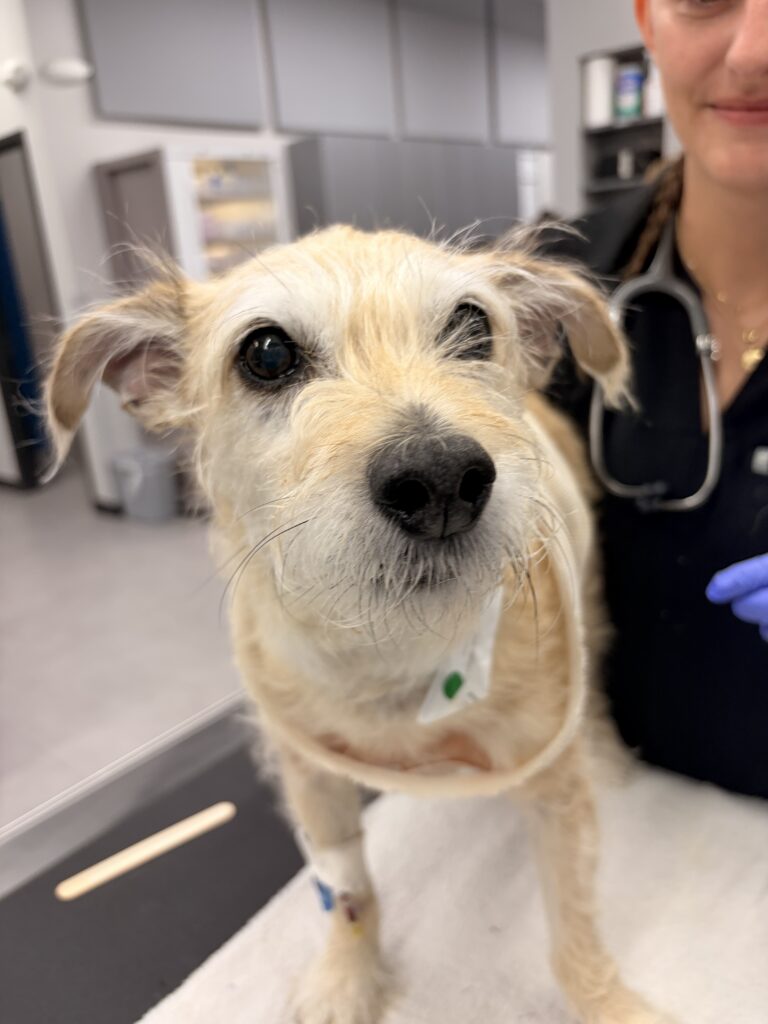
Herschel is an 11.5-year-old Terrier cross who initially presented in March for discomfort when being picked up. At that time, a chest mass was identified, though he was otherwise clinically stable at home, and the owners were appropriately concerned about surgical risk and timing.
By June, his clinical signs had progressed, including labored breathing as well as limping in a back limb. At that point, repeat CT imaging revealed a rather impressively sized cranial mediastinal mass. Prior testing performed by Dr. Cletzer indicated that the mass was most consistent with a thymoma (a benign, slow-growing mass) rather than lymphoma, which helped guide the treatment approach.
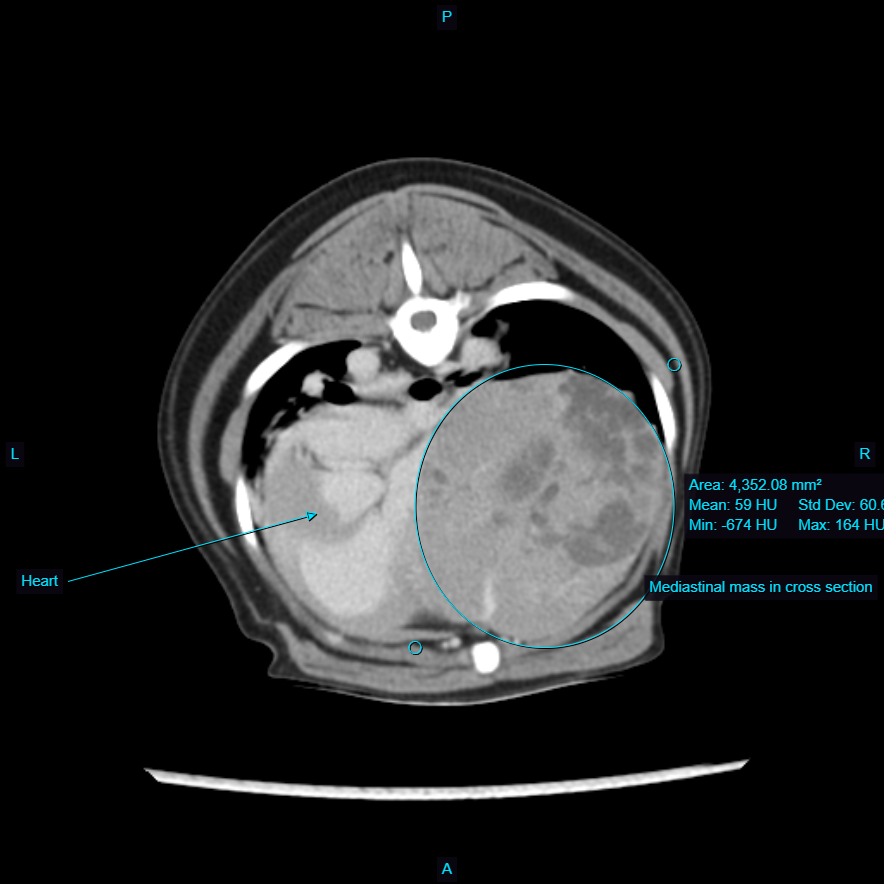
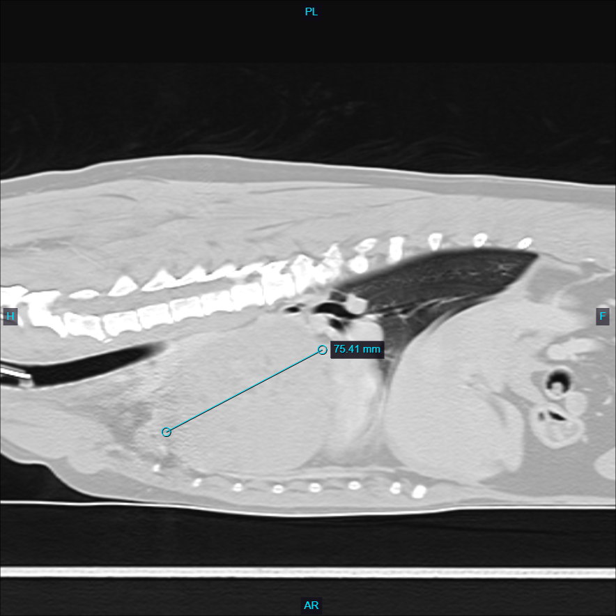
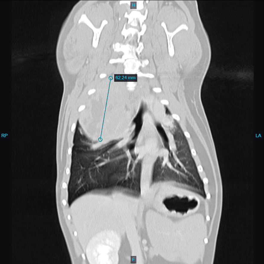
Herschel was taken to surgery, and the mass was successfully removed. Due to adhesions, a lung lobe also had to be removed as part of the procedure. He stayed one night in hospital after surgery and was discharged the following day, continuing to recover well at home.
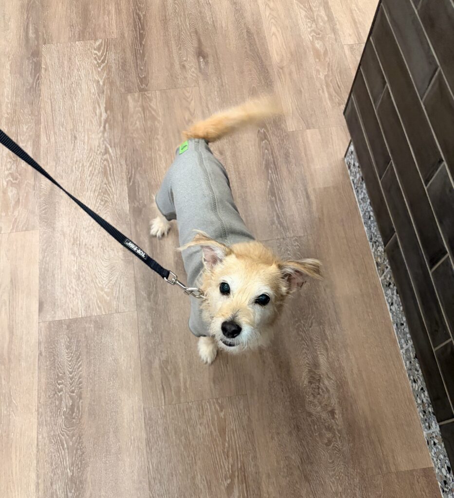
He was recently seen back for suture removal and presented in his cute recovery body suit, doing very well overall! His owner is very pleased with his recovery progress and outcome.
His case involved strong multidisciplinary collaboration, including diagnostic imaging and interpretation by Dr. Sepulveda (ultrasound, CT, and radiographs). Dr. Cletzer also performed important pre-operative testing to help differentiate lymphoma versus thymoma, which was critical in surgical planning and prognosis.